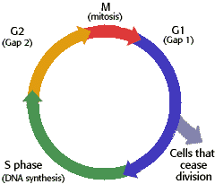
Monday, June 9, 2008

Cell Cycle Stages

Interphsae Mitosis

-----GAP 1 ----Prophase
-synthesize RNA -centromeres separate
-produce proteins -microtubule stretche
-Checkpoint: Everything ready for DNA synthesis?

-----GAP 0 (Cells that cease division) ----Prometaphase
-If cell too small -Nuclear membrane broken
-unfavorable environment
-then delay cell duplication ----Metaphase
-chromatids align on plate
-----Apoptosis
-p53 recognize damaged cell ----Anaphase
-if unable to repair damage -chromatids separate
- macrophage recognize and disassemble cell -separate to ends
- building blocks reused
----Telophase
-----S Phase -nuclear membrane reformed
-DNA synthesis
-All DNA replicated w/ protein molecules ----Cytokinesis
-cell pinches in two
-----Gap 2
-grow and produce proteins
-Checkpoint: Can proceed to mitosis to divide?

Mathematical Relationships


Stroke Volume (SV): Volume of blood being pumped by a ventricle in one contraction.
-Example: 70 ml
Heart Rate (HR)
The heart rate is the number of contractions (heart beats) in one minute. Two common ways of determining this are by placing two fingers on your wrist (radial artery), or on your neck (carotid artery).
-Example: 80 beats per minute (bpm)
Cardiac Output:
The cardiac output is the volume of blood being pumped by a ventricle in one minute. Logically, you can find this by multiplying how much blood is pumped in one contraction, by how many contractions there are in one minute.
-Stroke Volume multiplied by Heart Rate
-Example: 70 ml(SV) x 80 bpm(HR) = 5600 ml/minute
Systolic Pressure: Pressure of blood in artery when the heart contracts
-Example: 123 mm
Diastolic Pressure: Pressure of blood in artery while the heart relaxes in between beats
-Example: 81 mm
A systolic pressure of 123 mm and a diastolic pressure of 81 mm would give a blood pressure of 123/81 (systolic over diastolic) This is read as “123 over 81.”

Heart Rate Control


Atrial Contraction
Atrial contraction is the broad term for when the atria of the heart contract. This is regulated by the SA Node.
SA Node
The SA Node, or the Sinoatrial node, is a tissue that instigates the left and right atrium to contract by generating electrical impulses. The SA node is located along the right atrium of the heart. The tissue forces a contraction 60 to 100 times a minute.
Ventricular Contraction
Ventricular contraction is the broad term for when the ventricles of the heart are forced to contract. This comes after the atrial contraction.
AV Node
The AV Node, or the Atrioventricular node, is an area of tissue that delays ventricle contraction. It is located between the atria and ventricles of the heart. After a short delay from the atria contraction, the AV node instigates two bundle branches (Bundle of His) to contract the left and right ventricles.
Purkinje Fibers
The two bundle branches (Bundle of His) branch off into the Purkinje Fibers. These fibers spread across the bottom portion of the heart, instigating the heart’s “big” contraction. On an ECG, the biggest wave is caused by the Purkinje Fibers.


Effects of Diseases


Heart Attack – Sometimes, oxygen-supplying arteries for the heart become blocked up by plaque and platelets. The heart cannot receive oxygen. It cannot proceed in pumping blood and oxygen throughout the body, leading to death.
Atherosclerosis – In atherosclerosis, fatty substances, cholesterol, and other wastes can build up in arteries. If the plaque ruptures, the blood can clot, leading to a heart attack or a stroke. Then, the heart cannot receive oxygen, leading to death.
Stroke – A stroke is a disturbance in the blood vessels that give blood to the brain. This may be caused by a blockage in the blood vessels, or accumulation of blood in the skull. If the heart cannot pump blood to the brain due to blockage, this means death.
Hypertension – Hypertension is the elevation of blood pressure. High blood pressure can wear the heart out, leading to early death.
Heart Murmur – Heart murmurs are caused by the valves closing as blood moves through the heart. Heart murmurs themselves, are an irregular noise. Abnormal murmurs may imply a defect with the heart valves, infection, disease, or age.
Congestive Heart Failure – Congestive Heart Failue is where the heart cannot pump enough blood to all of the body’s organs. The heart pumps blood slowly, and the returning blood backs up, causing congestion in the tissues.

Passive and Active Transport


Passive and active transport are the two main means of transport in a cell. Passive transport requires no energy. Some examples of passive energy are diffusion, facilitated diffusion, and osmosis. In diffusion, liquids, gases, and solids can move in and out of a cell without energy. In facilitated diffusion, diffusion is occurred but with some type of carrier. Osmosis is diffusion for water. Passive transport always involves the movement of molecules from high concentration to low concentration. Active transport, on the other hand, always involves the movement of molecules from low concentration to high concentration. It requires energy in the form of ATP. Some examples of active transport are ion pumps, exocytosis, and endocytosis. Ion pumps are proteins that gather ions on one side of the membrane. In exocytosis, a vesicle holding material can break off the membrane and leave the cell. In endocytosis, a cell ingests a substance into the cell which then forms a vacuole.
http://www.accessexcellence.org/RC/VL/GG/ecb/ecb_images/12_04_passive_active_transport.jpg

Parts of Cells

Animal Plant


Eukaryotic - Prokaryotic

Eukaryotic

-Nucleus

-Membrane-bound organelles

-DNA enclosed

-Plants, animals, protists, fungi

-divides by mitosis

-goes through meisos in sexual reproduction

-large ribosomes


Prokaryotic

-No Nucleus
-No organelles
-Free-floating DNA
-Bacteria, archaea
-binary fission
-transfers DNA w/ conjugation
-fairly small ribosomes
-Fimbriae


Both

Both
-enclosed by plasma membrane
-contain
DNA
-have flagellum
-diverse in forms
-reproduce
-need energy
-perform same functions (like excretion_

http://www.cod.edu/people/faculty/fancher/ProkEuk.htm

http://earthlife.net/kingdom.html

http://life.umd.edu/classroom/bsci424/BSCI223WebSiteFiles/ProkaryoticvsEukaryotic.htm

http://www.cod.edu/people/faculty/fancher/EukaryoticCell.jpg


Cell Membrane


Membrane Parts

Function

Phospholipid Bilayer

Barrier for cell

Channel Protein

Channels certain molecules into cell

Carrier Protein

Moves molecules from high to low concentration

Cell Recognition Protein

Recognizes other cells which helps alert to foreign molecules

Receptor Protein

Connects internal and external environment of cell to sense surroundings

Enzymatic Protein

Produces enzymes to synthesize substances for cell

http://www.biologycorner.com/resources/cell_membrane.jpg

http://www.biologymad.com/cells/cellmembrane.htm

http://sun.menloschool.org/~cweaver/cells/c/cell_membrane/fluid_mosaic.jpg

http://en.wikipedia.org/wiki/Protein_Function_in_Cell_Membranes

Cell Parts and Functions

Cell Wall – The cell wall is a protective layer around a plant cell that gives support and controls cell size.


Cell Membrane – The cell membrane is a protective layer around the cytoplasm of a cell that is involved in diffusion and osmosis by its permeability.

Nucleus – The nucleus is the organelle that directs all cell activities and is the ‘director’ of the cell.

Endoplasmic Reticulum – The ER’s function is to store and transport molecules such as proteins throughout the cell. There are two types of ER: rough ER and smooth ER (lacking ribosomes).

Riboosomes – Ribosomes are the part of the cell that synthesize proteins. Some are embedded into the rough ER.

Mitochondria – The mitochondria can be considered the power factory of a cell because it creates energy from ATP. Also, it recycles proteins, fats, and carbohydrates, and is where cellular respiration takes place.

Chloroplast – The chloroplast is an organelle that is only in plant cells. It contains chlorophyll, which uses sunlight to produce energy (photosynthesis).

Vacuole – The vacuole stores water, digests food, removes waste, and releases water from unicellular organisms.

Cytosol – Cytosol is the fluid in the cell which the organelles are in. It’s mostly made of water and other floating molecules. Not to be confused with cytoplasm, cytosol itself does not contain organelles.

Lysosome – The lysosome is a sac with digestive enzymes that break up food molecules. It transports undigested food through the cell membrane and out of the cell.

Peroxisome – Peroxisomes remove toxic substances from the body such as hydrogen peroxide.

Golgi Apparatus – The Golgi Apparatus stores and packages proteins.

Cytoskeleton – The cytoskeleton shapes a cell and gives it strong support and protection.

http://upper.usm.k12.wi.us/academics/faculty/rheun/organelle.html
http://library.thinkquest.org/12413/structures.html
http://cellbio.utmb.edu/cellbio/lysosome.htm